blank plant cell diagram to label pdf
Plant cells, the foundational units of plant life, possess unique structures vital for survival and growth․
Understanding these components is greatly aided by utilizing diagrams, especially blank ones for interactive learning․
These PDF resources, readily available online, empower students and educators to visually explore and label cellular parts․
What are Plant Cells?
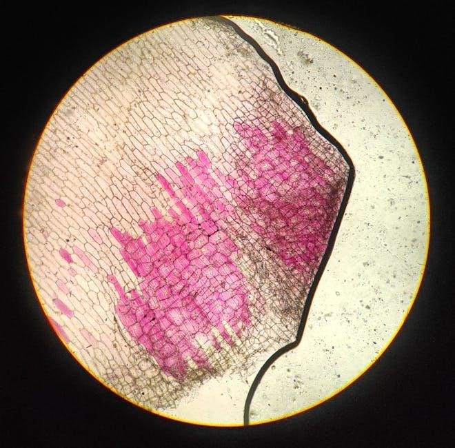
Plant cells are the basic building blocks of all plants, fundamentally differing from animal cells due to several key structures․ Most notably, they possess a rigid cell wall providing support and shape, alongside chloroplasts – organelles responsible for photosynthesis, the process of converting light energy into chemical energy․ These cells aren’t simply isolated units; they collaborate to form tissues, organs, and ultimately, the entire plant organism․
A blank plant cell diagram to label PDF serves as an invaluable educational tool․ It allows students to actively engage with the cell’s anatomy, reinforcing their understanding of each component’s function․ By identifying and labeling structures like the nucleus, vacuole, and cell membrane, learners solidify their knowledge in a practical and memorable way․ These diagrams often highlight the unique features distinguishing plant cells, fostering a deeper appreciation for the intricacies of plant biology․
Why Use a Labelled Diagram?
Utilizing a labelled diagram, particularly a blank plant cell diagram to label PDF, dramatically enhances the learning experience․ Visual representation simplifies complex biological concepts, making them more accessible and understandable․ Actively labeling the different parts – like the cell wall, chloroplasts, and nucleus – promotes deeper engagement and retention of information compared to simply reading text․
These diagrams aren’t merely for students; they’re valuable for educators too, serving as effective teaching aids․ A blank diagram encourages active recall and self-assessment․ The process of identifying and naming each structure reinforces understanding of its specific function within the cell․ Furthermore, readily available PDF formats offer convenience and portability, allowing for flexible learning both inside and outside the classroom, fostering a more comprehensive grasp of plant cell biology․

Key Components of a Plant Cell
Plant cells feature distinct parts – walls, membranes, nuclei, and cytoplasm – crucial for function․
Diagrams, especially PDF versions for labeling, illuminate these structures and their roles within the cellular system․
Cell Wall: Structure and Function
The cell wall is a rigid outer layer unique to plant cells, providing support, protection, and shape․ Composed primarily of cellulose, it differs significantly from animal cell structures․ Understanding its layered composition – the primary, middle lamella, and sometimes secondary walls – is key to comprehending plant cell function․
Diagrams, particularly those designed as blank plant cell diagrams to label in PDF format, are invaluable tools for visualizing these layers․ Students can actively learn by identifying and labeling the different wall components․ The cell wall’s porosity allows for the passage of water and nutrients, essential for plant life․ It also plays a crucial role in maintaining turgor pressure, contributing to plant rigidity and preventing wilting․ Utilizing a labelled diagram enhances comprehension of this vital structural element․
Cell Membrane: Controlling Entry and Exit
The cell membrane, present in all cells, acts as a selective barrier regulating the passage of substances in and out of the plant cell․ Composed of a phospholipid bilayer with embedded proteins, it controls transport, maintaining cellular homeostasis․ This dynamic structure is crucial for nutrient uptake, waste removal, and communication with the external environment․
Employing a blank plant cell diagram to label, often available as a PDF, allows students to pinpoint the cell membrane’s location and appreciate its role relative to other organelles․ Visualizing its position surrounding the cytoplasm reinforces understanding․ Diagrams highlight the membrane’s permeability and the function of transport proteins․ Interactive labelling exercises solidify knowledge of how this vital boundary controls cellular traffic, ensuring optimal plant cell function and survival․
Nucleus: The Control Center
The nucleus serves as the plant cell’s command center, housing the genetic material – DNA – organized into chromosomes․ It directs all cellular activities, including growth, metabolism, and reproduction․ This vital organelle is surrounded by a nuclear envelope, a double membrane with pores facilitating transport․ Within the nucleus, the nucleolus is responsible for ribosome synthesis․
Utilizing a blank plant cell diagram to label, frequently found as a downloadable PDF, provides a clear visual representation of the nucleus’s central location․ Students can accurately identify its components and understand its relationship to other organelles․ Labelling exercises reinforce the concept of the nucleus as the cell’s control hub, emphasizing its crucial role in coordinating all cellular processes for plant life and function․
Cytoplasm: The Cellular Environment
Cytoplasm comprises everything within the cell membrane, excluding the nucleus․ It’s a gel-like substance, primarily water, salts, and proteins, where organelles reside and cellular processes occur․ This dynamic environment facilitates biochemical reactions, nutrient transport, and waste removal, essential for cell survival․ The cytoplasm provides structural support and a medium for organelles to interact․
A blank plant cell diagram to label, often available as a free PDF download, effectively illustrates the cytoplasm’s expansive nature․ Students can visually appreciate how it fills the cell and surrounds all other components․ Labelling exercises using these diagrams reinforce understanding of the cytoplasm as the cell’s internal environment, crucial for supporting life functions and enabling interactions between organelles within the plant cell․

Organelles Within the Plant Cell
Plant cells contain specialized structures, called organelles, each with a unique role․
A blank plant cell diagram to label PDF helps visualize these components and their functions within the cell․
Chloroplasts: Photosynthesis Powerhouses
Chloroplasts are the sites of photosynthesis, the remarkable process where plants convert light energy into chemical energy in the form of sugars․ These organelles contain chlorophyll, a green pigment crucial for capturing sunlight․ A blank plant cell diagram to label PDF is an invaluable tool for students learning about these vital structures․
When using such a diagram, students can pinpoint the chloroplast’s location within the cell, often recognizing its distinctive shape – typically oval or disc-like․ Labeling exercises reinforce understanding of the internal structures like thylakoids and grana, where the light-dependent reactions occur․ Identifying these components on a diagram solidifies comprehension of how chloroplasts function as the energy producers of the plant cell, driving growth and sustaining life․ The PDF format allows for easy printing and repeated practice․
Mitochondria: Energy Production
Mitochondria are often called the “powerhouses” of the cell, responsible for generating most of the cell’s supply of adenosine triphosphate (ATP), used as a source of chemical energy․ Utilizing a blank plant cell diagram to label PDF provides a fantastic visual aid for understanding their structure and function․ Students can accurately place mitochondria within the cellular landscape․
Labeling exercises on these diagrams highlight key features like the inner and outer membranes, and the cristae – the folds that increase surface area for ATP production․ Recognizing these components reinforces the understanding of cellular respiration, the process where glucose is broken down to release energy․ A downloadable PDF allows for repeated practice and reinforces the crucial role mitochondria play in sustaining plant life, converting energy into a usable form․
Vacuole: Storage and Support
Vacuoles are large, fluid-filled organelles crucial for maintaining turgor pressure, storing nutrients, and waste products within plant cells․ A blank plant cell diagram to label PDF is an excellent tool for visualizing the vacuole’s prominent size and central location․ Students can practice accurately positioning and identifying this key component․
Labeling exercises using these diagrams emphasize the vacuole’s role in regulating cell volume and providing structural support․ The PDF format allows for repeated practice, solidifying understanding of how the vacuole contributes to plant rigidity and overall health․ Furthermore, it highlights the storage function, encompassing water, ions, and pigments․ Correctly identifying the vacuole on a diagram reinforces its importance in plant cell functionality and survival․
Ribosomes: Protein Synthesis
Ribosomes, essential for protein synthesis, are found throughout plant cells – both freely floating in the cytoplasm and bound to the endoplasmic reticulum․ Utilizing a blank plant cell diagram to label PDF allows students to pinpoint the often-numerous locations of these vital organelles․ This visual practice reinforces their understanding of ribosome distribution․

Labeling exercises with these PDF diagrams emphasize the ribosome’s critical role in translating genetic code into functional proteins․ Students learn to associate ribosomes with protein production, a fundamental process for plant growth and development․ The diagrams help visualize how ribosomes contribute to cellular functions․ Accurate placement on the diagram solidifies comprehension of their importance, and the PDF format enables repeated practice for mastery of this concept․
Endoplasmic Reticulum (ER): Transport Network
The Endoplasmic Reticulum (ER), a network of membranes, serves as the plant cell’s transport system․ A blank plant cell diagram to label PDF is invaluable for students to visualize the ER’s extensive, interconnected structure, distinguishing between rough ER (with ribosomes) and smooth ER․ This labeling practice reinforces understanding of its dual function․
Using these PDF diagrams, learners can accurately depict the ER’s role in synthesizing and transporting proteins and lipids․ The visual aid clarifies how the ER connects to the nucleus and Golgi apparatus, forming a continuous pathway for cellular traffic․ Identifying the ER on a diagram solidifies comprehension of its importance in cellular organization and function․ Repeated labeling with the PDF resource enhances retention and mastery of this complex organelle․
Golgi Apparatus: Processing and Packaging
The Golgi Apparatus functions as the plant cell’s processing and packaging center, modifying, sorting, and packaging proteins and lipids for transport․ A blank plant cell diagram to label PDF is crucial for students to accurately represent its stacked, flattened sacs, called cisternae․ This visual exercise reinforces understanding of its unique morphology․
Utilizing these PDF resources, learners can pinpoint the Golgi’s location near the ER and nucleus, illustrating its role in the secretory pathway․ Labeling the entry (cis) and exit (trans) faces on the diagram clarifies the directional flow of molecules․ Identifying vesicles budding off the Golgi emphasizes its packaging function․ Repeated practice with the PDF solidifies comprehension of this vital organelle’s role in cellular logistics and secretion․

Detailed Diagram Elements for Labelling
Detailed diagrams, often found as PDFs, require precise labelling of structures․ Accurate identification of organelles, like the nucleus and chloroplasts, enhances comprehension․
Identifying the Cell Wall Layers
Plant cell walls aren’t simple, uniform structures; they’re composed of distinct layers, each with a specific role․ When utilizing a blank plant cell diagram to label, accurately identifying these layers is crucial for understanding cell function․ The primary wall, the outermost layer, is relatively thin and flexible, allowing for growth․
Beneath this lies the middle lamella, a pectin-rich layer that cements adjacent cells together․ In some cells, a thicker, more rigid secondary cell wall is present, providing additional support and protection․ This layer often contains lignin, contributing to its strength․
PDF diagrams designed for labelling often highlight these layers with different textures or shading․ Students should focus on recognizing the relative thickness and position of each layer – primary, middle lamella, and secondary (if present) – to demonstrate a comprehensive understanding of cell wall architecture․ Correctly labelling these components showcases a grasp of plant cell structure and its implications for plant life․
Locating Chloroplasts within the Cell
Chloroplasts, the sites of photosynthesis, are key organelles within plant cells․ When working with a blank plant cell diagram to label, accurately positioning these structures is essential․ Unlike some organelles, chloroplasts aren’t uniformly distributed; they tend to be concentrated in cells of the leaf mesophyll, maximizing light capture․
On a diagram, chloroplasts appear as oval-shaped structures, often numerous within a single cell․ They are typically found towards the periphery of the cell, allowing sunlight to penetrate deeper tissues․ PDF resources for labelling frequently depict chloroplasts with internal structures like thylakoids and grana, prompting detailed observation․
Students should practice identifying the characteristic shape and location of chloroplasts to demonstrate understanding of their function․ Correct placement on the diagram reinforces the link between structure and photosynthetic activity, vital for plant survival and energy production․
Pinpointing the Nucleolus
The nucleolus, a prominent structure within the nucleus, is crucial for ribosome biogenesis․ When utilizing a blank plant cell diagram to label, accurately locating the nucleolus demonstrates understanding of its function․ It isn’t a membrane-bound organelle, appearing as a dense region within the nucleus itself․
On a typical diagram, the nucleolus appears as a darker, often spherical area inside the nucleus․ PDF resources designed for labelling often highlight its central position within the nuclear envelope․ Students should note its size relative to the overall nucleus; it’s usually quite noticeable․
Correctly identifying the nucleolus reinforces the understanding of gene expression and protein synthesis․ Labelling exercises using these diagrams help visualize the connection between genetic information and cellular machinery, essential for plant cell function and growth․
Recognizing the Vacuole’s Size and Shape
The vacuole is a defining feature of mature plant cells, often dominating the cell’s volume․ When working with a blank plant cell diagram to label, recognizing its substantial size is key․ Unlike animal cells with smaller vacuoles, plant cells typically possess one large central vacuole․
Its shape isn’t always perfectly regular; PDF diagrams may depict it as a large, irregular sac filling much of the cell’s interior․ This shape is dictated by the cell’s needs for storage, maintaining turgor pressure, and waste disposal․ Students should observe how the vacuole pushes other organelles towards the cell periphery․

Accurately labelling the vacuole reinforces understanding of its vital roles in plant cell homeostasis․ Diagrams help visualize its importance in maintaining cell rigidity and facilitating essential cellular processes․

Resources for Printable Diagrams (PDF)
Numerous online platforms offer printable, blank plant cell diagrams in PDF format, ideal for educational purposes and interactive labelling exercises for students․
Free Online Plant Cell Diagram PDFs
Accessing free, downloadable plant cell diagram PDFs is remarkably easy with a wealth of resources available online․ Several educational websites specialize in providing these materials, catering to various learning levels – from elementary school introductions to more detailed high school biology studies․ These PDFs typically feature clearly illustrated blank plant cell diagrams, specifically designed for students to practice labelling the different organelles and structures;
Popular search terms like “blank plant cell diagram to label PDF”, “printable plant cell worksheet”, and “plant cell outline diagram” yield numerous results․ Websites such as Science Notes and Biology Corner frequently offer free, high-quality diagrams․ Furthermore, many teachers and educational content creators share their resources on platforms like Teachers Pay Teachers, often including blank diagrams as part of larger lesson plans․ Remember to always preview the PDF to ensure it meets your specific needs regarding detail and complexity before printing or distributing it․
Educational Websites Offering Labelled Diagrams
Numerous educational websites provide both labelled and, crucially, blank plant cell diagrams ideal for student practice․ These resources often accompany comprehensive biology lessons, offering interactive elements and detailed explanations of each cellular component․ Sites like Biology LibreTexts and Khan Academy present visually engaging diagrams alongside informative text, fostering a deeper understanding of plant cell structure․
Beyond simple diagrams, some platforms offer virtual cell explorations, allowing students to ‘dissect’ a plant cell digitally․ While not directly PDF downloads, these interactive tools enhance learning․ Searching for “plant cell diagram” on websites like CK-12 Foundation and BBC Bitesize reveals a range of labelled diagrams that can serve as answer keys for blank diagram exercises․ These websites frequently provide downloadable worksheets, including options to create custom quizzes and assessments based on the plant cell structure․
Using Diagrams for Educational Purposes
Blank plant cell diagrams, particularly in PDF format, are invaluable tools for reinforcing learning in biology classrooms․ They encourage active recall and promote a deeper understanding of cellular structures beyond simple memorization․ Students can independently label organelles, solidifying their knowledge of each component’s function within the plant cell․
These diagrams facilitate differentiated instruction; teachers can tailor assignments based on student skill levels․ More advanced learners might label complex structures and explain their interactions, while others focus on core organelles․ PDF format ensures consistent presentation and easy printing for in-class activities or homework․ Utilizing diagrams also supports visual learners, offering a concrete representation of abstract concepts․ Furthermore, labelled diagrams serve as excellent study aids for quizzes and exams, enhancing retention and academic performance․

Tips for Effective Labelling

Effective labelling of a blank plant cell diagram (PDF) requires understanding each organelle’s role before applying labels; clarity and conciseness are key!
Understanding the Function Before Labelling
Before embarking on labelling a blank plant cell diagram – often found as a downloadable PDF – it’s crucial to grasp the function of each cellular component․ Simply memorizing names isn’t enough; understanding why a structure exists and what it does dramatically enhances the learning process․
For instance, knowing that chloroplasts are the sites of photosynthesis – converting light energy into chemical energy – makes identifying them on the diagram more meaningful․ Similarly, recognizing the cell wall’s role in providing structural support and protection clarifies its position and importance․
This functional understanding transforms the exercise from rote memorization to active learning․ It allows you to connect the structure to its purpose, solidifying your knowledge of plant cell biology․ Utilizing resources alongside the PDF, like tutorials and FAQs, can further aid in comprehending these vital functions before attempting to label the diagram accurately․
Using Clear and Concise Labels
When completing a blank plant cell diagram – frequently available as a PDF for printing – prioritize clarity and conciseness in your labelling․ Avoid lengthy descriptions; single words or short phrases are generally sufficient to identify each structure accurately․ For example, instead of “the organelle responsible for energy production,” simply label it “Mitochondria․”
Ensure your labels are neatly written or typed, and use lines that clearly point to the corresponding cellular component․ Ambiguous or overlapping labels can create confusion and detract from the diagram’s educational value․ Consistency is also key; use the same terminology throughout․
Remember, the goal is to demonstrate understanding, not to showcase elaborate handwriting․ A well-labelled diagram, utilizing precise and succinct terms, effectively communicates your knowledge of plant cell anatomy․